Pune – Minimally access brain surgery technique uses specialised endoscopes with high resolution video cameras to perform surgery of the brain. This technique can be used to diagnose and treat neurological conditions such as brain tumours, hydrocephalus (fluid build-up in the brain) and movement disorders. Traditional brain surgery requires removing a part of the skull to allow access to the brain, but in minimal access brain surgery, it usually involves making more precise small openings that minimize collateral damage to surrounding scalp, brain, blood vessels and nerves, making it painless and even sutureless.
“Minimal access brain surgery is not more or less risky than traditional surgery when performed with the purpose of making the patient’s life easier and secure. As long as the goal of the procedure is not compromised, it will always offer something extra than open surgery, such as a better quality of life through faster recovery and less scarring. Plus, the prospect of undergoing a surgery, which involves only a small opening or no obvious opening, is mentally more acceptable to patients who are already traumatised and scared of an impending brain surgery.”
In some cases, minimally access neurosurgery brings an entirely new facet of treatment to the force, for example, in the treatment of hydrocephalus. Hydrocephalus is a condition frequently encountered in clinical practice, in which there is an abnormal amount of cerebrospinal fluid (CSF) accumulation in the cranium. Traditionally, the treatment of hydrocephalus has been through correcting the overproduction of CSF or diverting the build-up away from the head by surgically placing tubes called shunts inside the brain ventricles and draining it into body cavities. Though effective, shunts are occasionally associated with serious complications, including infection, over-drainage or malfunction. Now using video guidance and no hardware, a surgeon can insert an endoscope through a small hole in the skull into the third ventricle of the brain. There a perforation is made in a membrane to restore normal flow of CSF. Though not all hydrocephalus patients are eligible for this approach, approximately 70 to 80 per cent of properly selected patients are successfully treated this way.
“Minimally invasive neurosurgery is a young field of just a few decades old, the world over. In India, its advent started perhaps in the last two decades and neurosurgeons here have whole-heartedly embraced this new technology and are exploring the numerous possibilities it throws up. Presently, endoscopic third ventriculostomy (like in the case of hydrocephalus), endoscopic removal of pituitary tumours and other skullbase tumors, endoscopic treatment of intra-ventricular tumours, intraparenchymal and ventricular bleeds etc are some of the brain lesions which are commonly managed by this technique.
Endoscopic neurosurgery continues to evolve with technical contributions from neurosurgeons around the world. These approaches are technically demanding; require specialized instrumentation and significant surgical practice. With accessibility to real time imaging and advanced image guided navigation system, neurosurgeons are able to monitor a patient’s progress and perform more complicated surgeries with accuracy and improved clinical outcomes.”
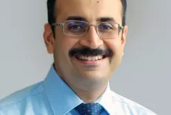
Dr Vishal Bhasme, Neuro surgeon, Lokmanya Hospital Pune
 Newspatrolling.com News cum Content Syndication Portal Online


